Structural basis for the prolonged photocycle of sensory rhodopsin II revealed by serial synchrotron crystallography.
Bosman, R., Ortolani, G., Ghosh, S., James, D., Norder, P., Hammarin, G., Ulfarsdottir, T.B., Ostojic, L., Weinert, T., Dworkowski, F., Tomizaki, T., Standfuss, J., Branden, G., Neutze, R.(2025) Nat Commun 16: 3460-3460
- PubMed: 40216733
- DOI: https://doi.org/10.1038/s41467-025-58263-x
- Primary Citation of Related Structures:
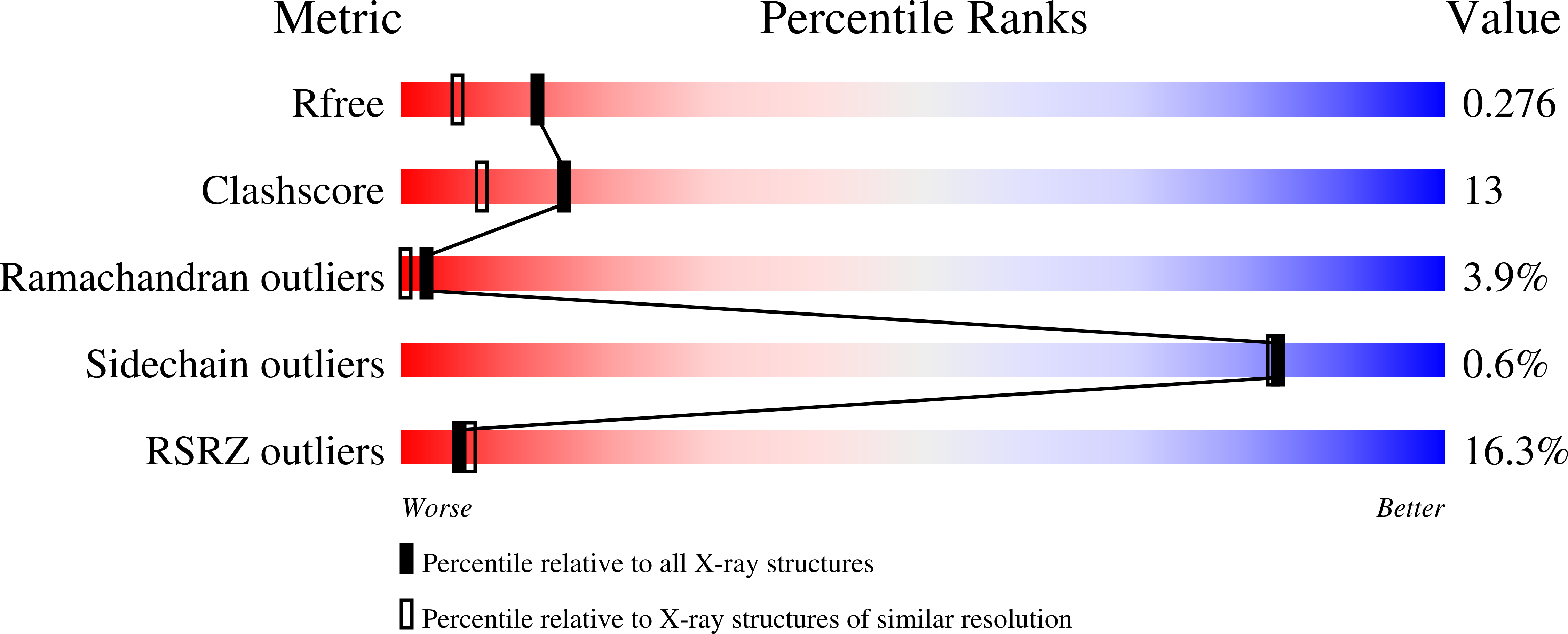
8PWG, 8PWI, 8PWJ, 8PWP, 8PWQ, 9H1W, 9H1X, 9H20 - PubMed Abstract:
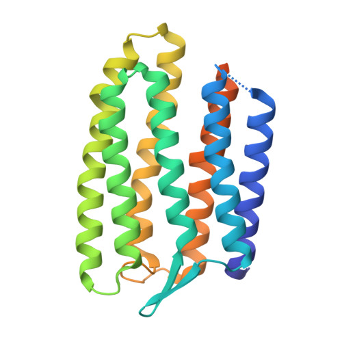
Microbial rhodopsins form a diverse family of light-sensitive seven-transmembrane helix retinal proteins that function as active proton or ion pumps, passive light-gated ion channels, and photosensors. To understand how light-sensing in archaea is initiated by sensory rhodopsins, we perform serial synchrotron X-ray crystallography (SSX) studies of light induced conformational changes in sensory rhodopsin II (NpSRII) from the archaea Natronomonas pharaonis, both collecting time-resolved SSX data and collecting SSX data during continuous illumination. Comparing light-induced electron density changes in NpSRII with those reported for bacteriorhodopsin (bR) reveals several common light-induced structural perturbations. Unlike bR, however, helix G of NpSRII does not unwind near the conserved lysine residue to which retinal is covalently bound and therefore transient water molecule binding sites do not arise immediately to the cytoplasmic side of retinal. These structural differences prolong the duration of the NpSRII photocycle relative to bR, allowing time for the light-initiated sensory signal to be amplified.
Organizational Affiliation:
Department of Chemistry and Molecular Biology, University of Gothenburg, G?teborg, Sweden.